Playing is Learning
The Specifics of the Frontal Eye
May 14th, 2015
| Ads | ||
|
Play the Challenge
|
||
|
A New Word is Coined A new Character is revealed A new Game is Afoot |
An Edutainment Adventure Based on Three Rounds of Investigations
|
  |
|
Welcome to the World of PROFESsee™by seeCOSM™ PROFESsee™ is my title. I am the perpetual learner, in pursuit of knowledge, wisdom and truth. I derived my name from professor |
 |
|
|
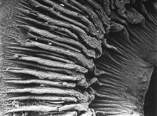
The human eye is one complex bit and without delving into too much biology, you will find it a handful. From your elementary school days where you learned that the eye perceives things as upside down, to when you started wondering whether that the teacher was right, the iris is a mystery, one that I really don’t care to decipher. One thing that is for sure though is that it is impossible to sneeze with your eyes open. I guess they would just pop out like missiles, iris and all. The iris of the human eye is the thin, circular colored part surrounding the pupil in the eye. It is usually called the ‘eye color’. Its primary function is controlling the size and diameter of the pupil thus regulating the amount of light that reaches the retina. The name iris is from the Greek goddess of the rainbow. It was named so because of its numerous colors. The optic cup part is the anterior two layers of an embryonic neuroectoderm structure where the iris develops. This cup is responsible for the production of the dilator and sphincter muscles of the iris. The iris of the human eye has two layers. The stroma - the front colored fibro vascular tissue and the colored epithelial cells located beneath the stroma. The stroma connects to the sphincter papillae muscle. This muscle is responsible for the circular motion contraction of the pupil. It is also connected to a set of dilator muscles. These muscles pull the iris to enlarge the pupil. The back part of the iris of the human eye is covered with a heavily pigmented epithelial layer. It is heavily colored due to the two cells thick of the epithelium. The front surface of the iris does not have an epithelium layer. The dilator muscles are found in the anterior surface of the iris. Due to its high colored content, it is able to block the amount of light reaching the retina. This is done through expansion and contraction of the muscles. The root of the iris is attached to the anterior ciliary and sclera body. It is divided into two major regions. The ciliary zone is the whole region extending to its origin at the body of the ciliary. The second region is the inner part called the pupillary zone. Can you find the answers related to the frontal eye? Image courtesy of: http://imgfave.com/search/part%20of%20the%20iris%20of%20a%20human%20eye |
||
Latest News / Events
E-mail [email protected]
The Professee™ Newsletter Beta
http://www.seecosm.com/
http://www.seecosm.com/